BACTERIA SHAPES AND GROUPING
ACTIVITY 1: BACTERIA SHAPES AND GROUPING
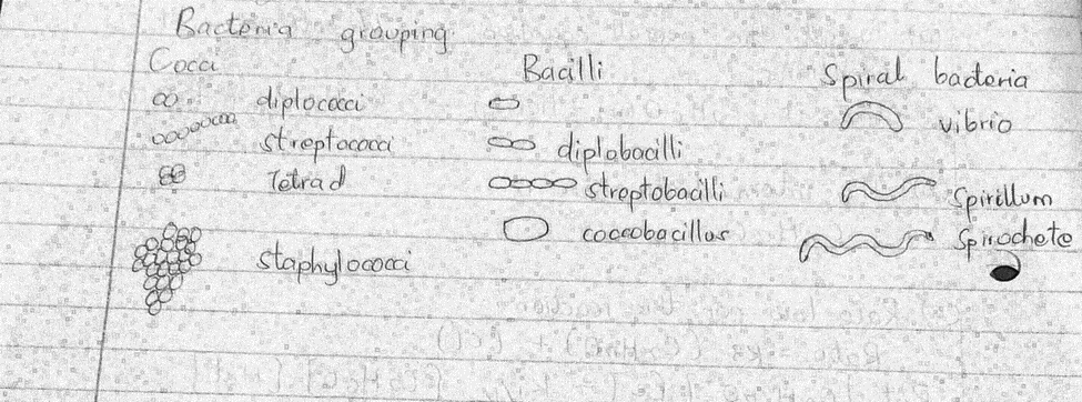
Q1. Obtain the slide labeled bacteria smear, alternatively, the slides maybe on display. Identify and draw the three distinct shapes of the organism using high power objective lens. Describe the appearance and grouping of the organism. Are the bacteria shown Gram-positive or Gram-negative? How do you know?

- Different features in bacteria are used in their classification. This includes their shape, number of flagella, and according to their harmfulness.
- According to their shapes, bacteria can be grouped into 3 main groups, that is, cocci, bacilli and spiral bacteria. Cocci include the round bacteria such as staphylococcus. Others appear round but hollow and are known as bacillus which includes streptobacillus. Some bacteria appear twisted to form long chains of different length and are known as spiral bacteria. This bacteria includes vibrio which are responsible for causing cholera.
- Gram staining is a lab procedure used to distinguish between different types of bacteria according to their staining ability. In this tests, gram positive bacteria usually gets stained whereas gram-negative retain the color. Harmful bacteria such as Clostridium botulinum which causes botulism are gram positive whereas Escherichia coli found in the environment is gram-negative.
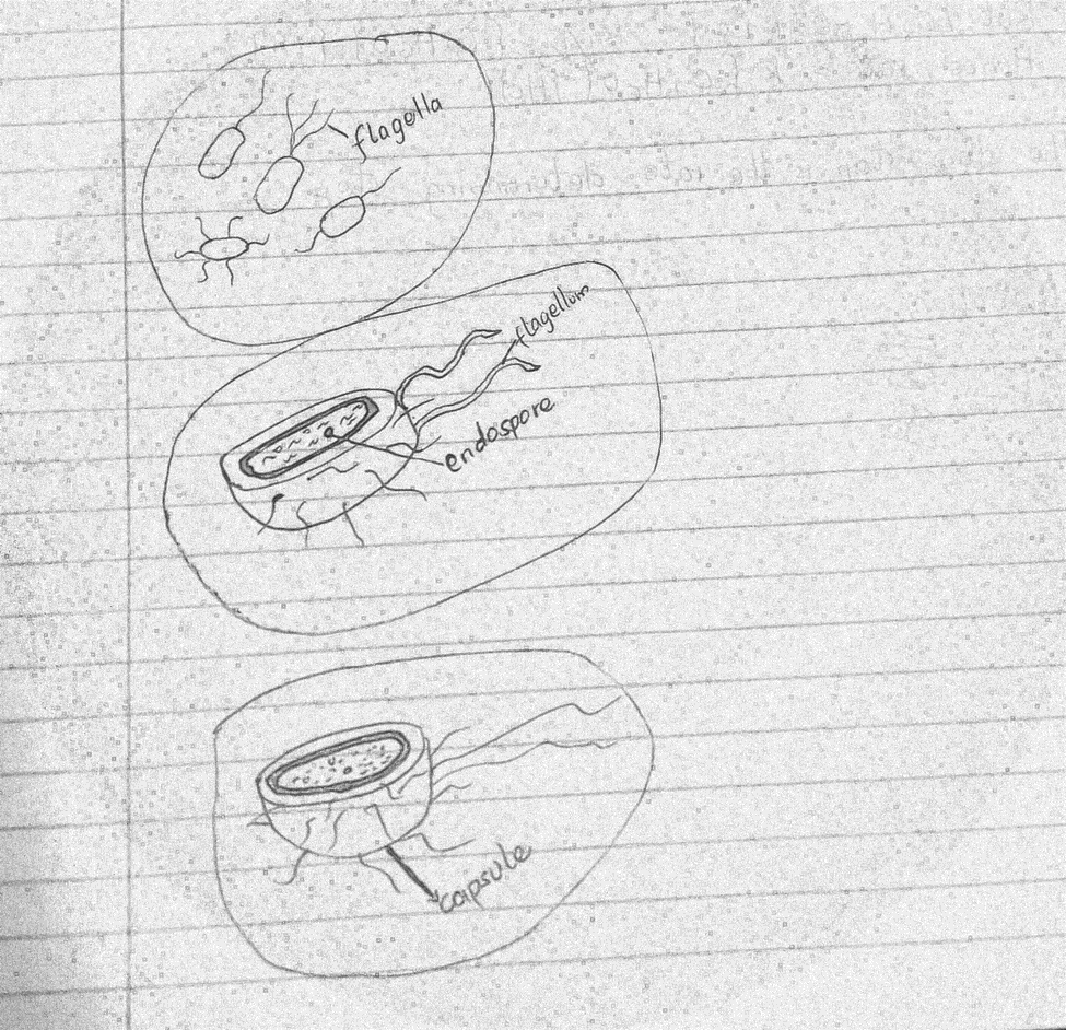
Q2. Draw the organism and describe its appearance. Make sure to label the flagella, capsule and endospore where appropriate

Flagella are locomotory structures normally found in bacteria. They are long helically shaped structures made up of the protein flagellin. It rotates in a clockwise and counterclockwise motion thus enabling bacteria to penetrate into many surfaces. The flagella attach to the bacterium through the envelope.
A capsule is a structure made up of a polysaccharide layer and is common in many bacteria. It lies on the outermost part of the bacteria and acts as a barrier thus protecting the bacteria from adverse conditions. It is a tough layer that cannot be removed easily and is responsible for causing many diseases.
An endospore is a tough, non-reproductive and dormant structure usually formed in some bacteria to allow it to navigate adverse environmental conditions. When conditions are not conducive, the bacteria reduce itself into a spore to allow it survive until when conditions resume.
ACTIVITY 2: View the following representatives of cyanobacteria. Identify unique structures and describe the type of colonies
| They are filamentous with solitary filaments Known for nitrogen-fixing abilities Colonies grow in filamentous clumps of multiple cell chains |
| Have specialized cells known as heterocyst Form flattened-rectangular colonies, that have one layer of cells. |
| Mostly found in symbiotic relationships with fungi. This type contains a lot of chlorophyll Its colonies are cocci shaped found in rocky places. |
| Are filamentous cyanobacteria found in freshwater? The filaments made of the thin gelatinous sheath Consists of filamentous blue-green colonies which are cylindrical |
| Are filamentous cyanobacteria This type consists of specialized cells known as heterocyst Forms flat-gelatinous colonies that are cylindrical, barrel-shaped up to form spherical shapes. |
POST-LAB QUESTIONS NAME:
- Differentiate between Bacteria and Archaea
Cell membrane in archaea is made up of pseudo peptidoglycan whereas in bacteria it is made up of either lipopolysaccharide or peptidoglycan. Archaea consists of three RNA whereas bacteria is made up of single RNA In archaea the main form of metabolism is methanogenesis whereas in bacteria it includes autotrophic, aerobic and anaerobic respiration, photosynthesis and fermentation.
- Streptococcus mutans is one of the bacteria responsible for dental plague formation and lacks an outer membrane outside its cell wall. Structurally and morphologically how would they look under the microscope? Illustrate the appearance.
S. mutans is a gram-positive bacterium, having a thick cell wall and retaining the gentle violet color during staining. It is a round bacterium when seen under a microscope. It isolates during staining to form round colonies which can be visualized under a microscope.

- In the search for extraterrestrial life, scientists expect that the most likely candidate would belong to one particular domain. Which domain would it be; Bacteria, Archaea or Eukarya? Explain why the expectation would be reasonable.
- The candidate will belong to archaea.
- This is because organisms in this domain have the ability to withstand harsh environments such as the extraterrestrial environment.
- These organisms are also said to play important roles in processes such as nitrogen fixation.
- Most archaea organisms are extremophiles which makes them suitable and easy to survive in an environment such as this.
- Below is a list of bacteria that causes disease in humans. Determine the clade, shape, and the associated disease of each bacterium.
- Bacterium
- Clade
- Shape
- Disease
- Chlamydia trachomatis
- Planctobacteria
- Rod/coccoid shaped
- Pelvic inflammatory disease
- Helicobacter pylori
- Campylobacteria
- Spiral shaped
- Peptic ulcers
- Clostridium botulinum
- Clostridia
- Rod-shaped
- Botulism
- Mycobacterium tuberculosis
- Clade 1
- Rod-shaped
- Tuberculosis
- Treponema pallidum
- Spirochaetes
- Helical
- Syphilis
- Yersinia pestis
- Gamma proteobacteria
- Rod-shaped
- Plague
- Haemophilus influenzae
- Gamma
- proteobacteria
- Rod-shaped
- Meningitis
- Why are typical bacteria and cyanobacteria classified together in the domain Bacteria? How do they differ?
- Both are classified in the same domain because they are prokaryotes. Both have a nucleus without a nuclear membrane, and lack membrane-bound organelles and therefore are grouped into the same domain. They possess a saprophytic mode of life. They both form resting spores. However, the two differ in the sense that cyanobacteria possess chlorophyll which most bacteria lack. Other differences include;
Bacteria Cyanobacteria In bacteria cell wall is made up of lipopolysaccharide and glycolipids. In Cyanobacteria it is made up of cellulose and pectin Most have flagella Flagella is absent here Spore formation in bacteria is endogenous In cyanobacteria, it’s not endogenous The mode of nutrition in bacteria may be autotrophic or heterotrophic In cyanobacteria, it is majorly autotrophic heterocyst may be found in bacteria Heterocyst are absent in cyanobacteria - Identify 3 benefits of bacteria to humans.
- Many bacteria are useful in the pharmaceutical industries and used in making drugs such as vaccines and antibiotics.
- Some bacteria are useful in the water and sewage companies as they are utilized to help in decomposing and degradation of harmful substances such as oil spills and other toxic
- wastes.
- Some bacteria are useful in food processing industries, such are Streptomyces are used in making food products such as yogurt, cheese, and alcoholic drinks.
- Describe 3 benefits of bacteria to the ecosystem.
- Bacteria are important decomposers. Are useful in the degradation of harmful substances in the environment
- Bacteria are environmental biosensors useful in sensing of toxic substances and thus help in cubing hazardous substances in the environment.
- Bioremediation is a process where selected bacteria are used to decompose harmful substances from industries and thus help to maintain the environment clean for healthy living.
- Search for an empirical or review article about a specific bacteria/ bacterial disease or a type of archaea. Summarize the article and include the relevance of the research. Cite the article using APA format.
- Helicobacter pylori
- H. pylori is a gram-negative spiral bacterium.
- This is a type of bacteria that infects the human stomach.
- It is responsible for stomach cancer that affects the mucosa-lymphoid tissue in the human stomach.
- It is responsible for stomach ulcers such as peptic and duodenal ulcers
- H. pylori attacks the lining that protects your stomach
- Ulcer symptoms may include;
- Be a dull pain that doesn’t go away
- Happen 2 to 3 hours after you eat
- Come and go for several days or weeks
- Happen in the middle of the night when your stomach is empty
- Go away when you eat or take medicines that reduce your stomach acid level (antacids)
- Interest in understanding the role of bacteria in stomach diseases was rekindled in the 1970s, with the visualization of bacteria in the stomachs of people with gastric ulcers.] The bacteria had also been observed in 1979, by Robin Warren, who researched it further with Barry Marshal in 1981. After unsuccessful attempts at culturing the bacteria from the stomach, they finally succeeded in visualizing colonies in 1982, when they unintentionally left their Petri dishes incubating for five days over the Easter weekend. In their original paper, Warren and Marshall contended that most stomach ulcers and gastritis were caused by bacterial infection and not by stress or spicy food, as had been assumed before.
- REFERENCES
- CY,Sheu BS,Wu JJ, Helicobacter pylori infection: An overview of bacterial virulence factors and pathogenesis. Biomedical journal. 2016 Feb [PubMed PMID: 27105595]
- Fischbach W,Malfertheiner P, Helicobacter Pylori Infection. Deutsches Arzteblatt international. 2018 Jun 22 [PubMed PMID: 29999489]